44 fluorescent labels and light microscopy
Light Microscope- Definition, Principle, Types, Parts, Labeled Diagram ... A light microscope is a biology laboratory instrument or tool, that uses visible light to detect and magnify very small objects and enlarge them. They use lenses to focus light on the specimen, magnifying it thus producing an image. The specimen is normally placed close to the microscopic lens. › us › enFluorophore selection | Thermo Fisher Scientific - US Photostability in buffer is a relative measure of the percentage of initial fluorescence intensity remaining following 30 seconds of continuous illumination using a 40x/1.4NA objective and a 100-watt Hg-arc lamp as the light source with samples mounted in phosphate buffer; high photostability in buffer is ideal for live-cell imaging and detection
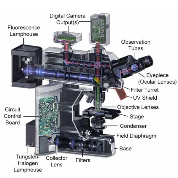
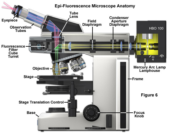
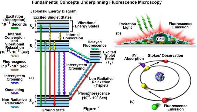
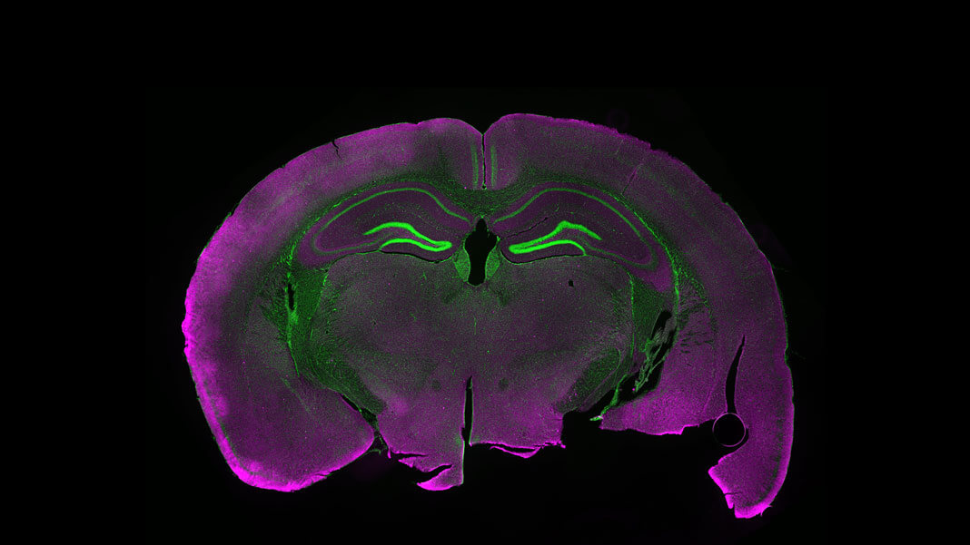
Fluorescence Microscopy - an overview | ScienceDirect Topics Fluorescence microscopy is a technique whereby fluorescent substances are examined in a microscope. It has a number of advantages over other forms of microscopy, offering high sensitivity and specificity. In fluorescence microscopy, the specimen is illuminated (excited) with light of a relatively short wavelength, usually blue or ultraviolet (UV).

Fluorescent labels and light microscopy
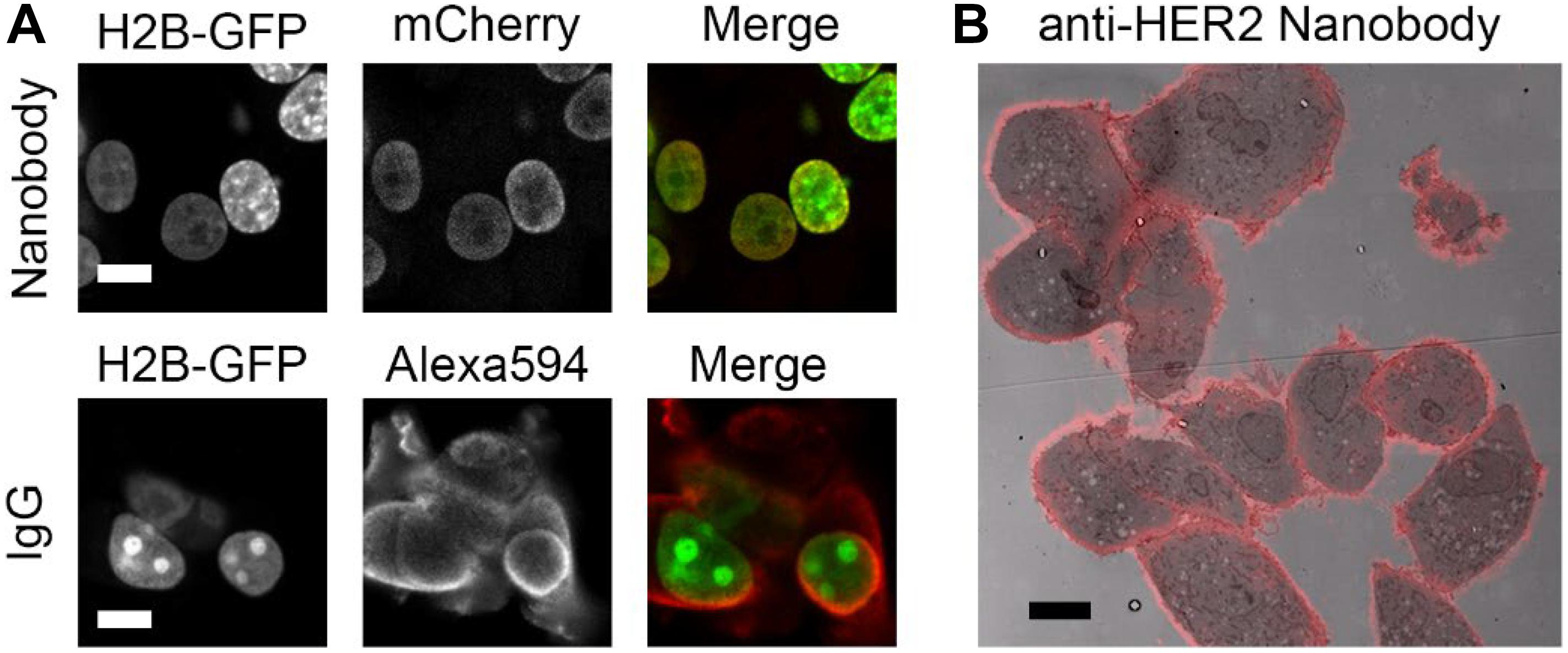
Fluorescent Dyes | Science Lab | Leica Microsystems In fluorescence microscopy there are two ways to visualize your protein of interest. Either with the help of an intrinsic fluorescent signal - by genetically linking a fluorescent protein to a target protein - or with the help of fluorescently labeled antibodies that bind specifically to a protein of interest. Fluorescent Labels - Neon Labels Inkjet/Laser | OnlineLabels® Our fluorescent label materials are bright, highly visible, neon colored labels with a permanent adhesive. The various five colors are ideal for signage, product labeling, organization, color coding and more. They have a matte finish that's great for printing logos, text, and designs and are both laser and inkjet printable for flexibility in ... Fluorescent Label - an overview | ScienceDirect Topics 14.3.2 Fluorescence quenching microscopy Fluorescence microscopy is a very common tool. Usually, fluorescent labels are used to brighten up the object of interest. However, the same strategy is not applicable for graphitic materials, such as graphite, graphene, GO or r-GO as they are strong quenchers of dye molecules [45].
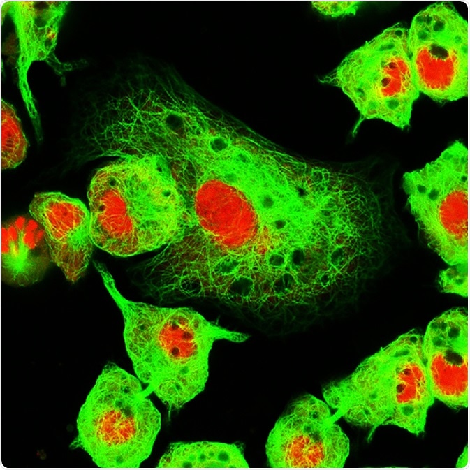
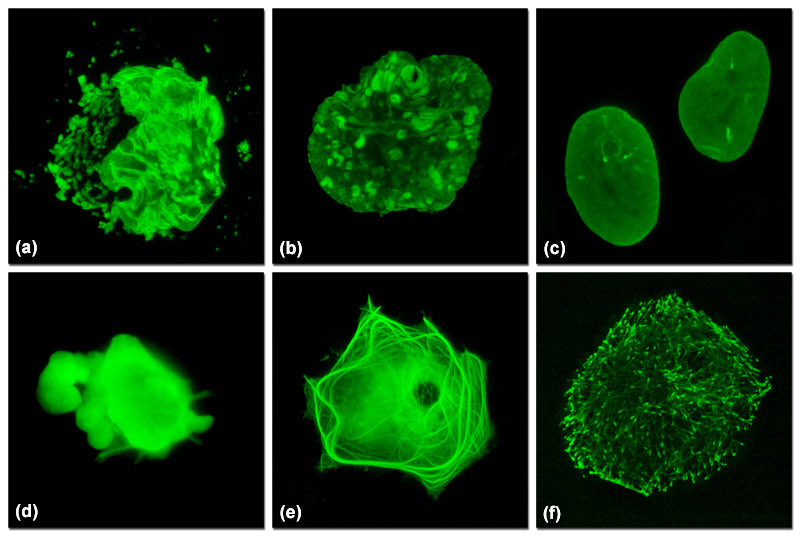
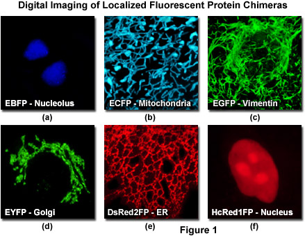
Fluorescent labels and light microscopy. Fluorescence in Microscopy | Science Lab | Leica Microsystems Fluorescence in Microscopy. Fluorescence microscopy is a special form of light microscopy. It uses the ability of fluorochromes to emit light after being excited with light of a certain wavelength. Proteins of interest can be marked with such fluorochromes via antibody staining or tagging with fluorescent proteins. Fluorescence Microscopy vs. Light Microscopy - News-Medical.net This means that fluorescent microscopy uses reflected rather than transmitted light. For example, a commonly used label is green fluorescent protein (GFP), which is excited with blue... Dots, Probes and Proteins: Fluorescent Labels for Microscopy and Imaging GFP now comes in 'flavors' including cyan, yellow and blue. Fluorescent proteins are useful for studying live cells and can be used as 'reporters' for studying gene expression. Using genetically modified plasmid and/or viral DNA, the target cells can be transfected with the plasmid which encodes both the fluorescent protein and a gene ... Light Sheet Fluorescence Microscopy | Nikon's MicroscopyU Light Sheet Fluorescence Microscopy (LSFM) is a general name for a constantly growing family of planar illumination techniques that have revolutionized how optical imaging of biological specimens can be performed. Fundamentally, LSFM techniques are made possible by decoupling the illumination and detection optical pathways, allowing for novel illumination strategies that optimize the photon ...
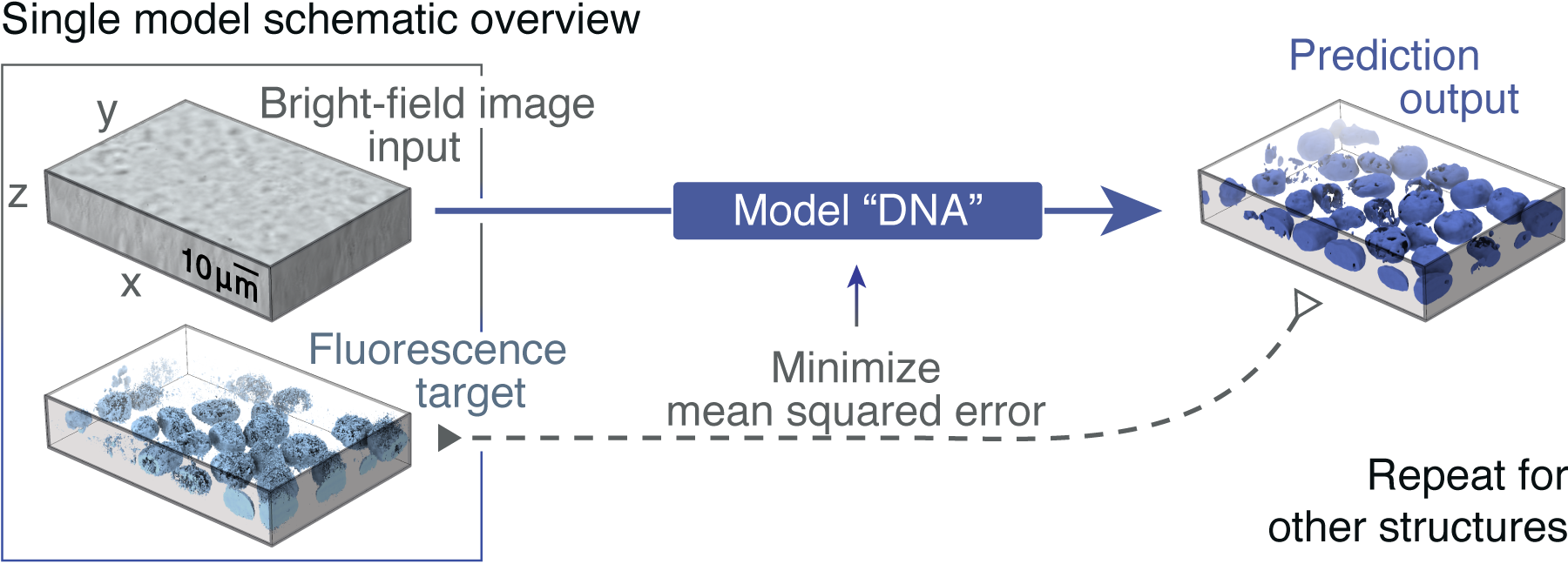
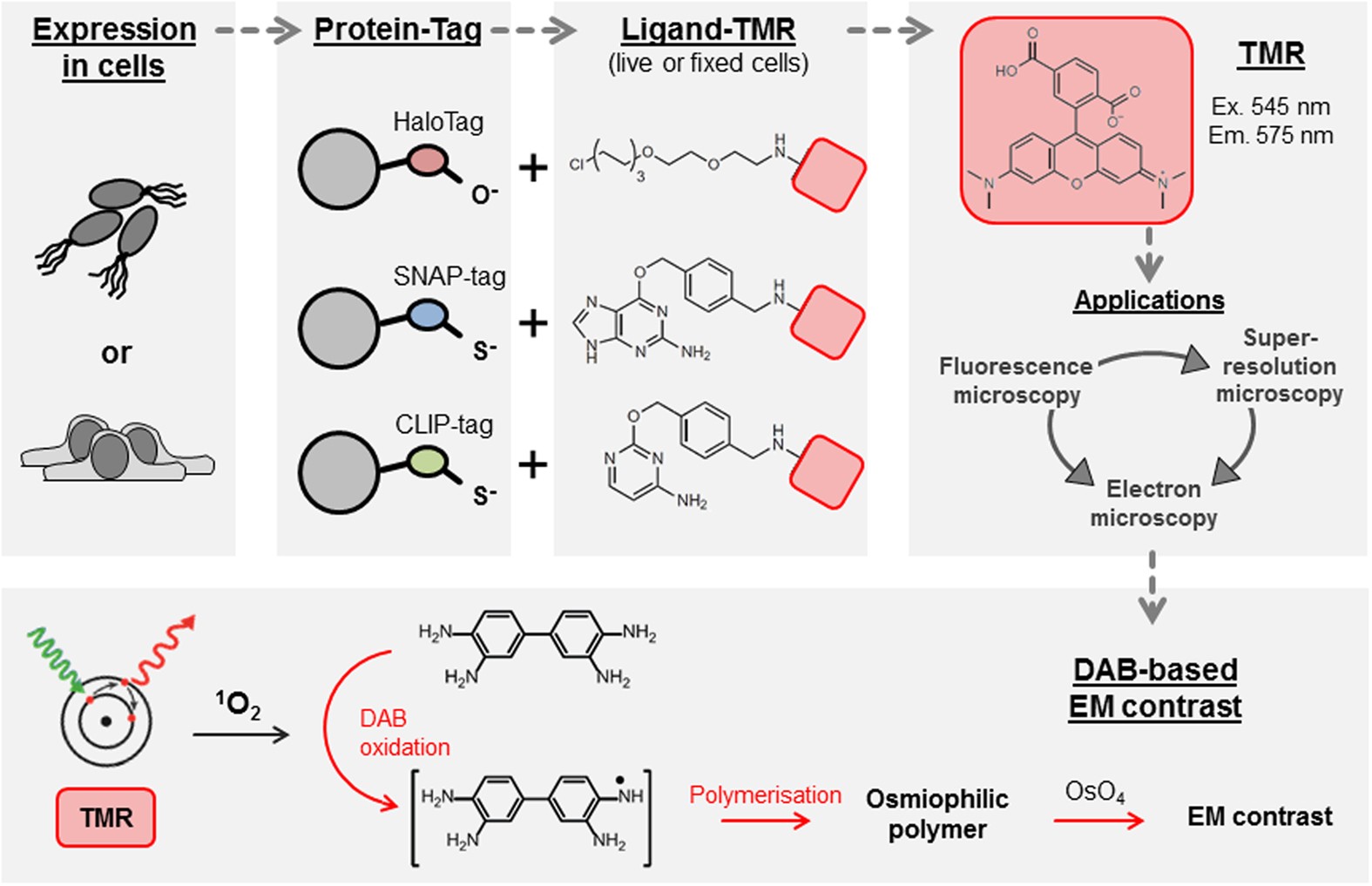
Different Ways to Add Fluorescent Labels - Thermo Fisher Scientific Using fluorescence provides greater contrast compared to viewing your samples with brightfield microscopy alone. Labeling various targets with separate fluorescent colors allows you to visualize different structures or proteins within a cell in the same experiment. In Silico Labeling: Predicting Fluorescent Labels in Unlabeled Images Microscopy is a central method in life sciences. Many popular methods, such as antibody labeling, are used to add physical fluorescent labels to specific cellular constituents. ... (ISL), reliably predicts some fluorescent labels from transmitted-light images of unlabeled fixed or live biological samples. ISL predicts a range of labels, such as ... › applications › fretBasics of FRET Microscopy | Nikon’s MicroscopyU The first fluorescent protein biosensor was a calcium indicator named cameleon, constructed by sandwiching the protein calmodulin and the calcium calmodulin-binding domain of myosin light chain kinase (M13 domain) between enhanced blue and green fluorescent proteins (EBFP and EGFP). In the presence of increasing levels of intracellular calcium ... Live Cell Imaging | Solutions | Leica Microsystems 01.06.2022 · Live‐cell imaging is mainly performed with fluorescence microscopy. Widefield microscopy, providing flexible excitation and fast acquisition, is typically used to visualize cell dynamics and development over long times. Confocal microscopy is typically used to study subcellular dynamic events. Multiphoton microscopy allows excitation with ...
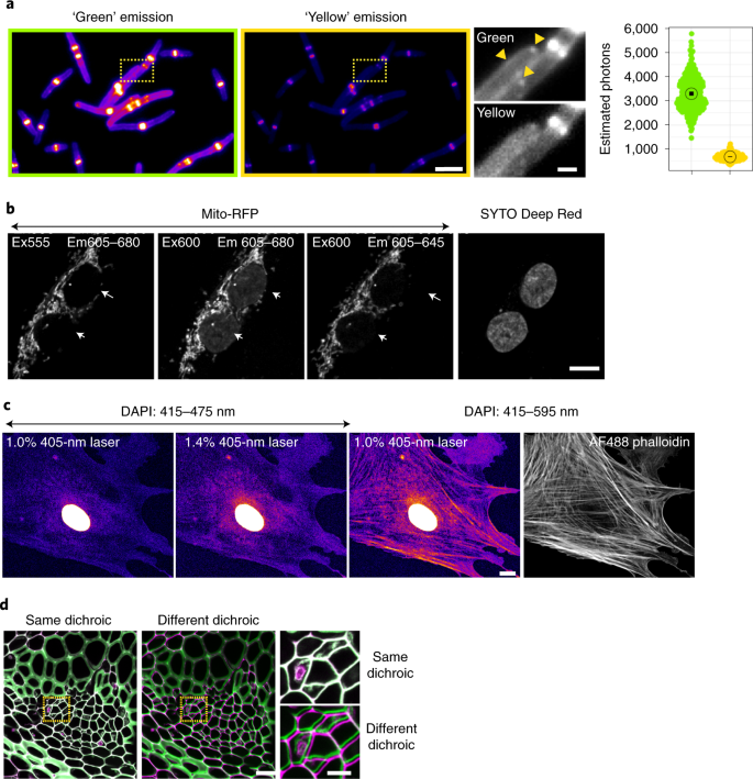
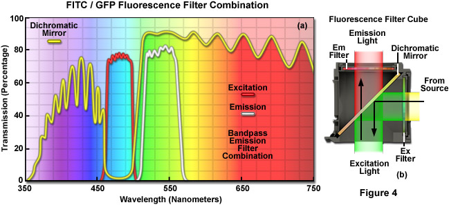
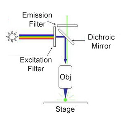
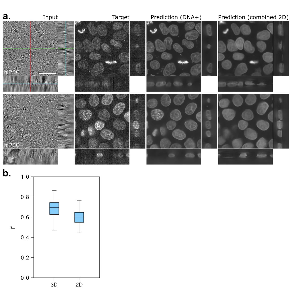
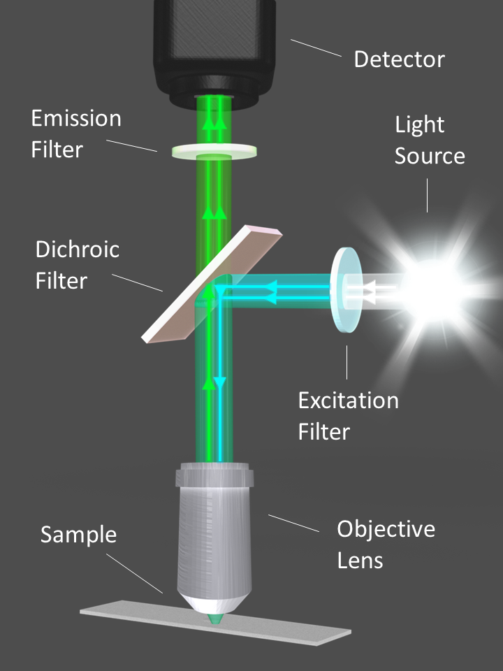
Introduction to Fluorescence Microscopy | Nikon's MicroscopyU It is important to note that fluorescence is the only mode in optical microscopy where the specimen, subsequent to excitation, produces its own light. The emitted light re-radiates spherically in all directions, regardless of the excitation light source direction. Label-free prediction of three-dimensional fluorescence images from ... Label-free prediction of three-dimensional fluorescence images from transmitted-light microscopy Understanding cells as integrated systems is central to modern biology. Although fluorescence microscopy can resolve subcellular structure in living cells, it is expensive, is slow, and can damage cells. Why would scientists use fluorescent labels or dyes when using a light ... What is the advantage of fluorescence microscopy over light microscopy? Advantages of Fluorescence Microscope Fluorescence microscopy is the most popular method for studying the dynamic behavior exhibited in live-cell imaging. This stems from its ability to isolate individual proteins with a high degree of specificity amidst non-fluorescing ... Fluorescent Labeling - What You Should Know - PromoCell Fluorescence microscopy allows the identification of cells and cellular components and the monitoring of cell physiology with high specificity. Fluorescence microscopy separates emitted light from excitation light using optical filters. The use of two indicators also allows the simultaneous observation of different biomolecules at the same time.
Fluorescent Label - an overview | ScienceDirect Topics 14.3.2 Fluorescence quenching microscopy Fluorescence microscopy is a very common tool. Usually, fluorescent labels are used to brighten up the object of interest. However, the same strategy is not applicable for graphitic materials, such as graphite, graphene, GO or r-GO as they are strong quenchers of dye molecules [45].
Fluorescent Labels - Neon Labels Inkjet/Laser | OnlineLabels® Our fluorescent label materials are bright, highly visible, neon colored labels with a permanent adhesive. The various five colors are ideal for signage, product labeling, organization, color coding and more. They have a matte finish that's great for printing logos, text, and designs and are both laser and inkjet printable for flexibility in ...
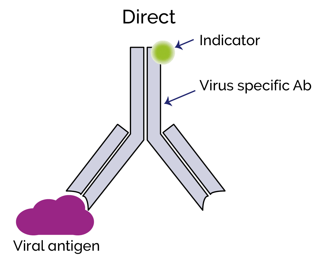
Fluorescent Dyes | Science Lab | Leica Microsystems In fluorescence microscopy there are two ways to visualize your protein of interest. Either with the help of an intrinsic fluorescent signal - by genetically linking a fluorescent protein to a target protein - or with the help of fluorescently labeled antibodies that bind specifically to a protein of interest.














Post a Comment for "44 fluorescent labels and light microscopy"