41 chlamydomonas diagram with labels
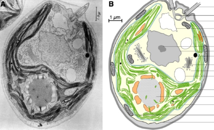
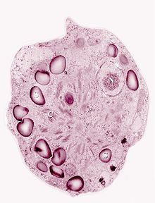
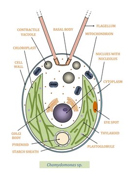
Chlamydomonas - Wikipedia Drawings of Chlamydomonas caudata Wille. [1] Cross section of a Chlamydomonas reinhardtii cell Light micrograph of Chlamydomonas with two flagella just visible at bottom left Chlamydomonas globosa, again with two flagella just visible at bottom left Use this labeled diagram of a chlamydomonas cell to - Course Hero Use this labeled diagram of a Chlamydomonas cell to address the following two questions. 32. Which of the following statements correctly identifies aspects related to photosynthesis and/or respiration? 1. Acetyl CoA is most often found in G. 2. NADPH accumulates in C. 3. ATP is found in F. 4.
Draw a neat labelled diagram. Chlamydomonas - Biology Draw a neat labelled diagram. Chlamydomonas . Maharashtra State Board HSC Science (General) 11th. Textbook Solutions 9073. Important Solutions 19. Question Bank Solutions 5548. Concept Notes & Videos 486. Syllabus. Advertisement Remove all ads. Draw a neat labelled diagram. ...

Chlamydomonas diagram with labels
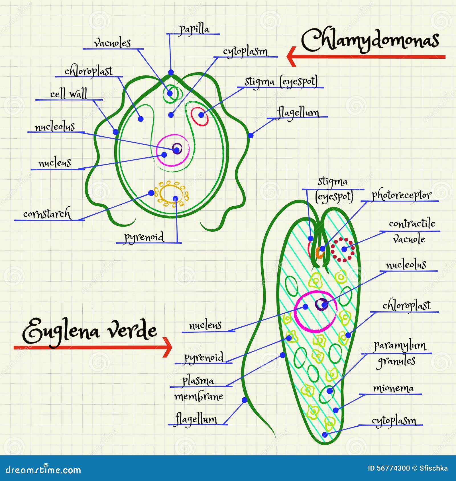
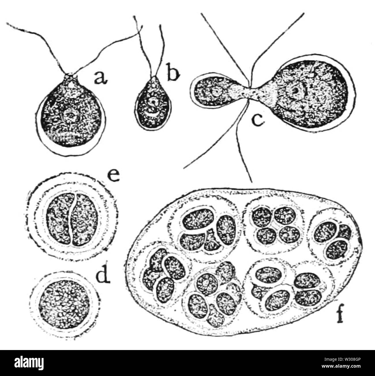
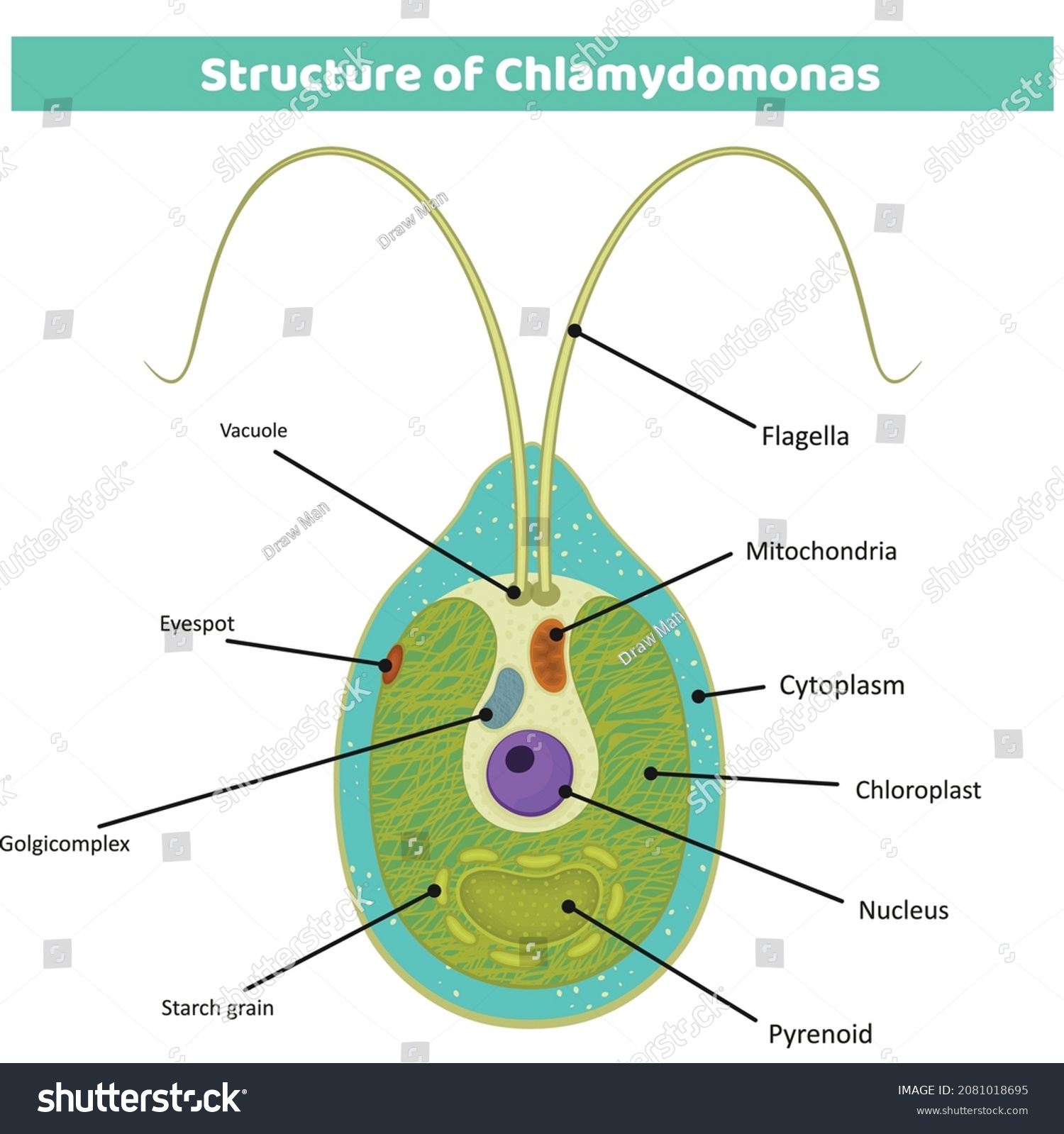
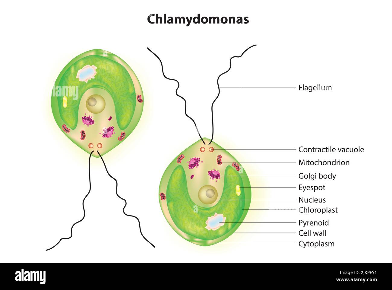
Structure of Chlamydomonas (With Diagram) | Chlorophyta In this article we will discuss about the structure of chlamydomonas with the help of suitable diagrams. Chlamydomonas is unicellular, motile green algae. The thallus is represented by a single cell. It is about 20 p,-30|i in length and 20 µ in diameter. The shape of thallus can be oval, spherical, oblong, ellipsoidal or pyriform. Use this labeled diagram of a chlamydomonas cell to Use this labeled diagram of a Chlamydomonas cell to address the following two questions. 32. Which of the following statements DOES NOT correctly match a function or component with its labelled feature? A. Channelrhodopsin can be found in C. B. Life Cycle of Chlamydomonas (With Diagram) - Biology Discussion Each daughter cell develops cell wall, flagella and transforms into zoospore (Fig. 6). The zoospores are liberated from the parent cell or zoosporangium by gelatinization or rupture of the cell wall. The zoospores are identical to the parent cell in structure but smaller in size. The zoospores simply enlarge to become mature Chlamydomonas.
Chlamydomonas diagram with labels. › doi › 10Life cycle and functional genomics of the unicellular red ... In the unicellular green alga Chlamydomonas reinhardtii, the BELL-related (GSP1) or KNOX (GSM1) gene is expressed only in mating-type–plus or mating-type–minus gametes, respectively, and the two proteins heteromerize, trigger nuclear and other organellar fusions between the two mating types , and activate diploid gene expression after mating . algae diagram class 8|algae drawing class 8|chlamydomonas diagram algae diagram class 8|algae drawing class 8|chlamydomonas diagramHi friends, In this video we will learn to draw draw algae diagram #algaedrawing... Diagram Of Chlamydomonas With Label / Queensland Lungfish Heart ... Answer for draw a labeled diagram of chlamydomonas. Shipping a package with ups is easy, as you can print labels for boxes, paste them and even schedule a pickup. Well labelled diagram of chlamydomonas. Jun 1, 2021 | hits: Creating your own labels is easy. This article details this process for you. Biology Laboratory Manual 12th Edition [12nbsped.] … Figure 3.1 The size of cells and their contents. This diagram shows the size of human skin cells, organelles, and molecules. In general, the diameter of a human skin cell is about 20 micrometers (µm), of a mitochondrion is 2 µm, of a ribosome is 20 nanometers (nm), of a protein molecule is 2 nm, and of an atom is 0.2 nm.
Morphology of Chlamydomonas (With Diagram) | Algae - Biology Discussion In this article we will discuss about the external morphology of chlamydomonas. Also learn about its Neuromotor Apparatus, Electron Micrograph, Palmella-Stage with suitable diagram. 1. The organism is an unicellular alga (Fig. 11). 2. The thallus is spherical to oblong in shape but some species are pyriform or ovoid. ADVERTISEMENTS: 3. Development of spirulina for the manufacture and oral delivery of ... Mar 21, 2022 · b, Diagram of primer pairs for PCR genotyping. Amplification of LHA and RHA includes one priming site (MP1 and MP4) present only in the spirulina genome at the target locus. Eye Diagram With Labels and detailed description - BYJUS Iris is the coloured part of the eye and controls the amount of light entering the eye by regulating the size of the pupil. The lens is located just behind the iris. Its function is to focus the light on the retina. The optic nerve transmits electrical signals from the retina to the brain. Pupil is the opening at the centre of the iris. › microorganisms-friend-and-foeMicroorganisms: Friend and Foe Class 8 Extra Questions ... Oct 11, 2019 · Pull out a gram or bean plant from the field. Observe its roots. You will find round struc¬tures called root nodules on the roots. Draw a diagram of the root and show the root nod¬ules. Answer: Question 2. Collect the labels from the bottles of jams and jellie on the labels. Answer: Do it yourself. Question 3. Visit a dcotor.
What Is Chlamydomonas? Explain With Diagram - Biology QA - BYJU'S Explain With Diagram Chlamydomonas is a unicellular, motile freshwater species and a genus of green algae. They are oval, spherical or slightly cylindrical in shape. Chlamydomonas is widely distributed in freshwater or in wet soil and is generally found in a habitat rich in ammonium salt Find Jobs in Germany: Job Search - Expat Guide to Germany Browse our listings to find jobs in Germany for expats, including jobs for English speakers or those in your native language. Chloroplast - Wikipedia A chloroplast / ˈ k l ɔːr ə ˌ p l æ s t,-p l ɑː s t / is a type of membrane-bound organelle known as a plastid that conducts photosynthesis mostly in plant and algal cells.The photosynthetic pigment chlorophyll captures the energy from sunlight, converts it, and stores it in the energy-storage molecules ATP and NADPH while freeing oxygen from water in the cells. The ATP and NADPH … Structure of Volvox (With Diagram) | Chlorophyta - Biology Discussion The cells of Volvox colony are Chlamydomonas type. Every cell has its own mucilage sheath (Fig. 1 B). The mucilage envelope of colony appears angular due to compression between cells. The cells are connected to each other through cytoplasmic strands. In some species of Volvox the cytoplasmic connections or strands are not present.
algae diagram | algae drawing | how to draw algae diagram ... algae diagram | algae drawing | how to draw algae diagram | chlamydomonas diagram | class 8 scienceHi friends, welcome to my Om Art Channel,In thi...
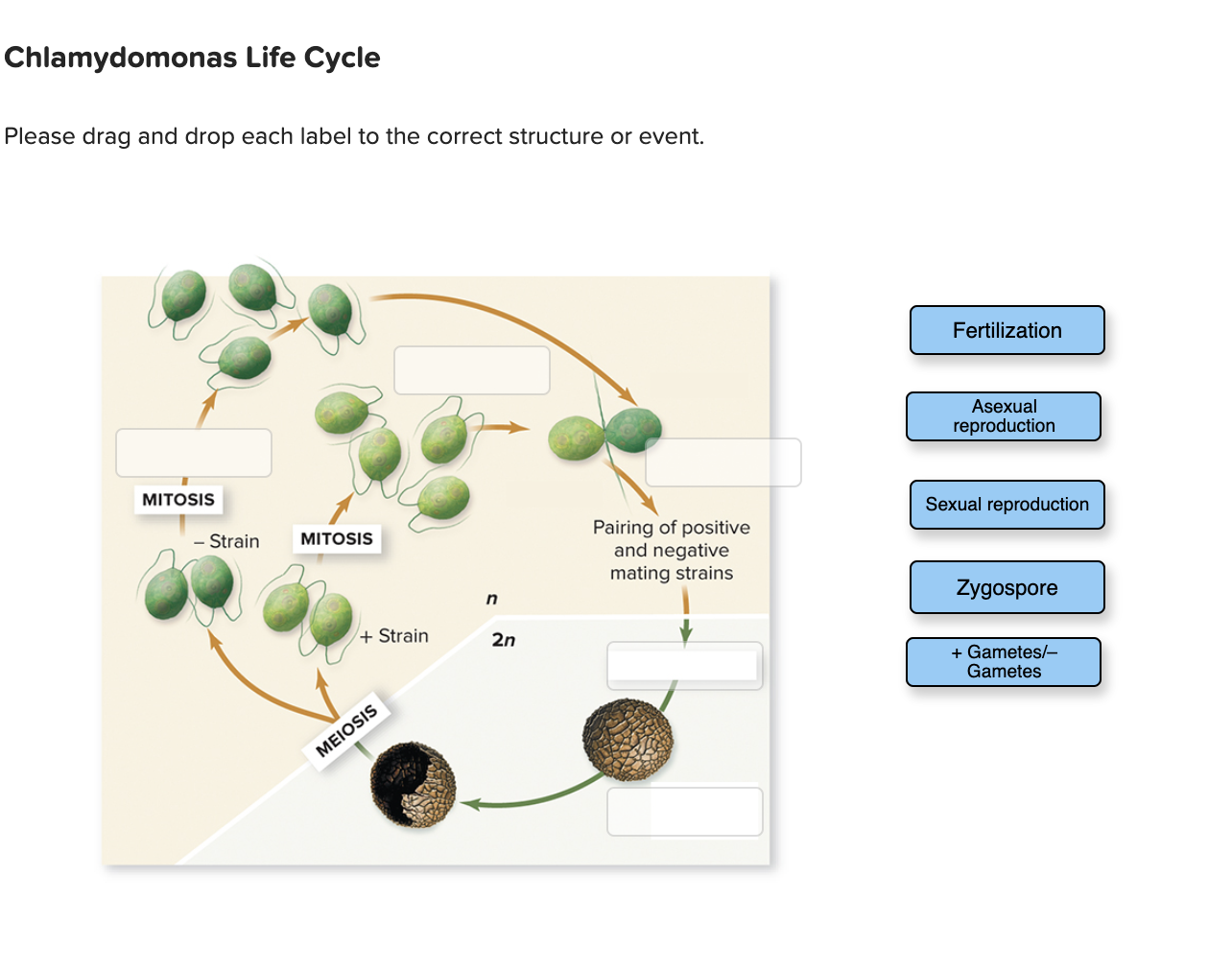
Structure of Chlamydomonas (With Diagram) | Genetics - Biology Discussion In this article we will discuss about the structure of chlamydomonas (explained with labelled diagram). The unicellular green alga Chlamydomonas is haploid with a single nucleus, a chloroplast and several mitochondria (Fig. 9.3). It can reproduce asexually as well as sexually by fusion of gametes of opposite mating types (mt + and mt - ).
issuu.com › cupeducation › docsCambridge IGCSE Biology Coursebook (third edition) - Issuu Jun 09, 2014 · Here are some points to bear in mind when you label a diagram. ♦♦ Use a ruler to draw each label line. ♦♦ Make sure the end of the label line actually touches the structure being labelled ...
Cambridge IGCSE Biology Coursebook (third edition) - Issuu Jun 09, 2014 · Here are some points to bear in mind when you label a diagram. ♦♦ Use a ruler to draw each label line. ♦♦ Make sure the end of the label line actually touches the structure being labelled ...
dokumen.pub › campbell-biology-12th-edition-12Campbell Biology, 12th Edition [12nbsped.] 9780135988046 ... In Chapter 12, the cell cycle figure (Figure 12.6) now includes cell images and labels describing the events of each phase. Unit 3 GENETICS Chapters 13–17 incorporate changes that help students grasp the more abstract concepts of genetics and their chromosomal and molecular underpinnings.
Chlamydomonas: Structure, Classification, and Characteristics Chlamydomonas is a genus of 325 species of unicellular green algae. The flagellates can be found living in droplets of water in freshwater, seawater, stagnant water, and even within moist soil. Chlamydomonas are studied as model creatures thanks to their unique flagellar movements and physiology.
Higher Education Support | McGraw Hill Higher Education Learn more about McGraw-Hill products and services, get support, request permissions, and more.
en.wikipedia.org › wiki › ChloroplastChloroplast - Wikipedia A chloroplast / ˈ k l ɔːr ə ˌ p l æ s t,-p l ɑː s t / is a type of membrane-bound organelle known as a plastid that conducts photosynthesis mostly in plant and algal cells.The photosynthetic pigment chlorophyll captures the energy from sunlight, converts it, and stores it in the energy-storage molecules ATP and NADPH while freeing oxygen from water in the cells.
Campbell Biology, 12th Edition [12nbsped.] 9780135988046 In Chapter 12, the cell cycle figure (Figure 12.6) now includes cell images and labels describing the events of each phase. Unit 3 GENETICS Chapters 13–17 incorporate changes that help students grasp the more abstract concepts of genetics and …
Campbell Biology, Third Canadian Edition (3rd Edition) [Third … Label the part of the diagram that represents the most recent common ancestor of frogs and humans. Alternative Forms of Tree Diagrams Fishes Frogs Chimps Lizards Chimps Humans Figure 6.32 Visualizing the Scale of the Molecular Machinery in a Cell, p. 132 Fishes Frogs 3 How many sister taxa are shown in these two trees? Identify them. 4
Chlamydomonas Diagram ️draw chlamydomonas, labeled science diagram# ... This video will be very useful for students to draw the structure of Chlamydomonas very easily.Thanks for watching and subscribe to the channel for drawing#...
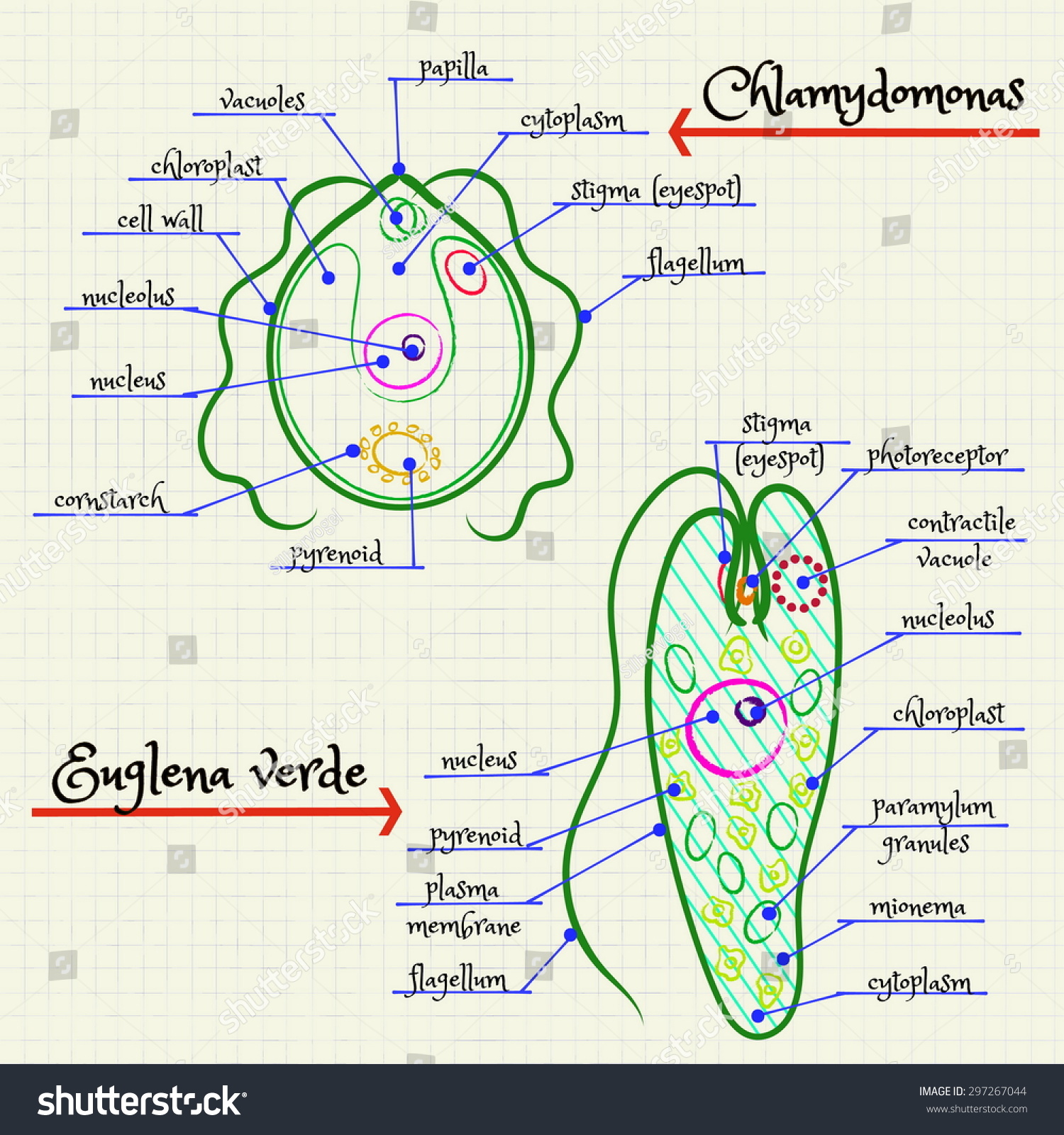
Diagram Of Chlamydomonas Cell / Solved Label The Parts Of The ... The anterior end of cytoplasm contains two . I tell you about how can we draw labelled diagram of chlamydomonas in . Biological drawings of protista, structure of chlamydomonas, resources for biology education by d g mackean. I tell you about how can we draw labelled diagram of chlamydomonas in .
Microorganisms: Friend and Foe Class 8 Extra Questions Oct 11, 2019 · Pull out a gram or bean plant from the field. Observe its roots. You will find round struc¬tures called root nodules on the roots. Draw a diagram of the root and show the root nod¬ules. Answer: Question 2. Collect the labels from the bottles of jams and jellie on the labels. Answer: Do it yourself. Question 3. Visit a dcotor.
Chlamydomonas Diagram drawing CBSE || easy way || Labeled Science ... These algae are found all over the world, in soil, fresh water, oceans, and even in snow on mountaintops. More than 500 different species of Chlamydomonas have been described, but most scientists...
Lehninger principles of biochemistry 6th edition pdf - Academia.edu Carbohydrates are the most abundant organic compounds in the plant world. They act as storehouses of chemical energy (glucose, starch, glycogen); are components of supportive structures in plants (cellulose), crustacean shells (chitin), and connective tissues in animals (acidic polysaccharides); and are essential components of nucleic acids (D-ribose and 2-deoxy-D …
› de › jobsFind Jobs in Germany: Job Search - Expat Guide to Germany ... Browse our listings to find jobs in Germany for expats, including jobs for English speakers or those in your native language.
› articles › s41587/022/01249-7Development of spirulina for the manufacture and oral ... Mar 21, 2022 · b, Diagram of primer pairs for PCR genotyping. Amplification of LHA and RHA includes one priming site (MP1 and MP4) present only in the spirulina genome at the target locus.
Describe the structure of chlamydomonas with neat labelled diagram ... Describe the structure of chlamydomonas with neat labelled diagram. micro organisms; class-8; Share It On Facebook Twitter Email. 1 Answer +1 vote . answered Oct 30, 2020 by Naaji (56.8k points) selected Oct 30, 2020 by Jaini . Best answer ...
Diagram Of Chlamydomonas With Label / Coloring Page With Structure Of ... Answer for draw a labeled diagram of chlamydomonas. It is oblong or pyriform in shape. Learn to make custom labels of your own. Click here to get an answer to your question ️ draw a labelled diagram of chlamydomonas. Well labelled diagram of chlamydomonas. To keep reading this answer, download the app. This article details this process for ...
Life Cycle of Chlamydomonas (With Diagram) - Biology Discussion Each daughter cell develops cell wall, flagella and transforms into zoospore (Fig. 6). The zoospores are liberated from the parent cell or zoosporangium by gelatinization or rupture of the cell wall. The zoospores are identical to the parent cell in structure but smaller in size. The zoospores simply enlarge to become mature Chlamydomonas.
Use this labeled diagram of a chlamydomonas cell to Use this labeled diagram of a Chlamydomonas cell to address the following two questions. 32. Which of the following statements DOES NOT correctly match a function or component with its labelled feature? A. Channelrhodopsin can be found in C. B.
Structure of Chlamydomonas (With Diagram) | Chlorophyta In this article we will discuss about the structure of chlamydomonas with the help of suitable diagrams. Chlamydomonas is unicellular, motile green algae. The thallus is represented by a single cell. It is about 20 p,-30|i in length and 20 µ in diameter. The shape of thallus can be oval, spherical, oblong, ellipsoidal or pyriform.













Post a Comment for "41 chlamydomonas diagram with labels"