38 structure of the heart with labels
Human Heart - Anatomy, Functions and Facts about Heart - BYJUS To know more about the human heart structure and function, or any other related concepts such as arteries and veins, ... Practice your understanding of the heart structure. Drag and drop the correct labels to the boxes with the matching, highlighted structures. Instructions to use: Hover the mouse over one of the empty boxes. One part in the image gets highlighted. Identify the … Human Heart Models | Heart Anatomy Models | Vitality Medical Heart Anatomy Models offer precise detail of the human heart and its structures to facilitate learning. The heart anatomy model is cast from durable elasticated plastic to resemble the natural position and scale of the human heart. 3B Scientific manufacturers a variety of life-size and 2-times life-size multi-dimensional models with dissection ...
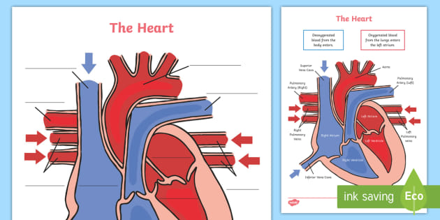
The Human Heart Labeling Worksheet (Teacher-Made) - Twinkl The human heart is a muscle made up of four chambers, these are: Two lower chambers - the left and right ventricles. It's also made up of four valves - these are known as the tricuspid, pulmonary, mitral and aortic valves. With this heart diagram without labels, you can familiarise your students with all the correct terms and help them ...

Structure of the heart with labels
Heart - Collection Page | AnatomyTOOL 3D model of the normal heart and lungs with numbered English labels. From a collaboration of Universities of Leiden, Delft and Groningen. 3D photogrammetry of plastinated opened normal heart A 3D photogrammetry of a plastinated opened normal heart by University of Maastricht. Video of Beating Heart Structure of the Heart | The Franklin Institute The heart consists of four chambers: two atria on the top and two ventricles on the bottom. Looking at the Valentine's Day heart, the two rounded humps at the top are rounded like the top of a lower-case "a." The bottom is shaped like a "v." Feel it working What else is inside your heart? Heart: Anatomy and Function - Cleveland Clinic The parts of your heart are like the parts of a house. Your heart has: Walls. Chambers (rooms). Valves (doors). Blood vessels (plumbing). Electrical conduction system (electricity). Heart walls Your heart walls are the muscles that contract (squeeze) and relax to send blood throughout your body.
Structure of the heart with labels. Heart Labeling Quiz: How Much You Know About Heart Labeling? Here is a Heart labeling quiz for you. The human heart is a vital organ for every human. The more healthy your heart is, the longer the chances you have of surviving, so you better take care of it. Take the following quiz to know how much you know about your heart. Questions and Answers 1. What is #1? 2. What is #2? 3. What is #3? 4. What is #4? Heart Diagram with Labels and Detailed Explanation - Collegedunia The heart is located under the ribcage, between the lungs and above the diaphragm. It weighs about 10.5 ounces and is cone shaped in structure. It consists of the following parts: Heart Detailed Diagram Heart - Chambers There are four chambers of the heart . The upper two chambers are the auricles and the lower two are called ventricles. Chapter 19: The Heart Flashcards | Quizlet •Allows heart to beat without friction, gives it room to expand and resists excessive expansion •Parietal pericardium-tough outer, fibrous layer of connective tissue-inner serous layer •Visceral pericardium (a.k.a. epicardium of heart wall)-serous lining of sac turns inward at base of heart to cover the heart surface Chapter 19: The Heart Flashcards | Quizlet •Allows heart to beat without friction, gives it room to expand and resists excessive expansion •Parietal pericardium-tough outer, fibrous layer of connective tissue-inner serous layer •Visceral pericardium (a.k.a. epicardium of heart wall)-serous lining of sac turns inward at base of heart to cover the heart surface
Anatomy | Label the Heart Diagram | Quizlet Right Atrium Receives deoxygenated blood from the body Right Ventricle Pumps deoxygenated blood to the lungs Left Atrium Chamber that receives oxygenated blood from the pulmonary veins Left Ventricle Pumps oxygenated blood into the aorta Aorta Largest artery in the body Superior Vena Cava Human Heart - Diagram and Anatomy of the Heart - Innerbody Chambers of the Heart The heart contains 4 chambers: the right atrium, left atrium, right ventricle, and left ventricle. The atria are smaller than the ventricles and have thinner, less muscular walls than the ventricles. The atria act as receiving chambers for blood, so they are connected to the veins that carry blood to the heart. How to Draw the Internal Structure of the Heart (with Pictures) - wikiHow To draw the internal structure of a human heart, follow the steps below. Part 1 Finding a Diagram 1 To find a good diagram, go to Google Images, and type in "The Internal Structure of the Human Heart". Find an image that displays the entire heart, and click on it to enlarge it. 2 Find a piece of paper and something to draw with. PDF Label the heart - Science Learning Hub Title: Label the heart Author: Science Learning Hub, The University of Waikato Created Date: 6/16/2017 1:02:16 PM
Heart Blood Flow | Simple Anatomy Diagram, Cardiac Circulation ... - EZmed Step 2 involves the left atrium, the chamber of the heart that receives oxygenated blood from the lungs via the pulmonary veins. 3. Mitral Valve Step 3 involves the mitral valve. During diastole, when the heart is relaxed and filling with blood, the oxygenated blood from the left atrium will flow to the left ventricle. Nutritionist Pro™ | Diet Analysis, Food Label, Menu Creation ... Designed and managed by registered dietitians for your comprehensive nutrition analysis needs. From food labels to menus to recipe calculations, Nutritionist Pro™ makes all your food science needs a simple and streamlined process. Since 1982 over 1,000,000 have relied on the Nutritionist Pro™ family of products. Carbohydrates | American Heart Association Apr 16, 2018 · Carbohydrates are either called simple or complex, depending on the food’s chemical structure and how quickly the sugar is digested and absorbed. The type of carbohydrates that you eat makes a difference – Foods that contain high amounts of simple sugars, especially fructose raise triglyceride levels. Ch. 19 Circulatory System- heart Flashcards | Quizlet Place the labels in order denoting the flow of blood through the pulmonary circuit beginning with the right atrium and ending in the left atrioventricular valve. The first and last structures are given. Right atrium 1. tricuspid valve 2. right ventricle 3. pulmonary valve 4. pulmonary trunk 5. pulmonary artery 6. lungs 7. pulmonary vein
Heart Diagram with Labels and Detailed Explanation - BYJUS Diagram of Heart. The human heart is the most crucial organ of the human body. It pumps blood from the heart to different parts of the body and back to the heart. The most common heart attack symptoms or warning signs are chest pain, breathlessness, nausea, sweating etc. The diagram of heart is beneficial for Class 10 and 12 and is frequently ...
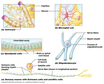
Nervous system - BIOLOGY4ISC
PDF Heart Structure - Indiana The heart is an organ about the size of a fist. It is made of muscle and pumps blood through the body. Tube-like structures called blood vessels carry blood through the body and heart. The heart and blood vessels make up the cardiovascular system. Structure of the Heart The heart has four chambers: two upper chambers call
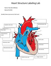
Lab Heart Structure Labeling. - Heart Structure Labeling Lab Name Hannah Kingsley Date Time 11: ...
Eating for a healthy heart - Baker This may help to improve your overall heart health. Unhealthy ‘saturated and trans’ fats should be limited. This fact sheet will help you with: choosing healthy fats and sources; practical ways to include this into your diet; reducing unhealthy fats in your diet; how to read nutrition food labels. Download fact sheet

A labelled diagram of the heart - Document in A Level and IB Human Biology
SGLT2 inhibitor - Wikipedia The differences in the structures is relatively small. The general structure includes a glucose sugar with an aromatic group in the β-position at the anomeric carbon. In addition to the glucose sugar moiety and the β-isomeric aryl substituent the aryl group is composed of a diarylmethylene structure.

Inspirierend Heart Diagram With Labels Gcse
Heart Health | Heart Attack Prevention | Bayer® Aspirin TO HELP PREVENT ANOTHER HEART ATTACK. A doctor-directed aspirin regimen helps keep your blood flowing. Along with other heart-healthy choices, it can reduce your risk of having another heart attack. Learn About Aspirin's Benefits. Aspirin is not appropriate for everyone, so be sure to talk to your doctor before you begin an aspirin regimen.
Heart Diagram Labelling Activity
The Anatomy of the Heart, Its Structures, and Functions - ThoughtCo The heart is the organ that helps supply blood and oxygen to all parts of the body. It is divided by a partition (or septum) into two halves. The halves are, in turn, divided into four chambers. The heart is situated within the chest cavity and surrounded by a fluid-filled sac called the pericardium. This amazing muscle produces electrical ...

SGLT2 inhibitor - Wikipedia SGLT2 inhibitors, also called gliflozins or flozins, are a class of medications that modulate sodium-glucose transport proteins in the nephron (the functional units of the kidney), unlike SGLT1 inhibitors that perform a similar function in the intestinal mucosa.The foremost metabolic effect of this is to inhibit reabsorption of glucose in the kidney and therefore lower blood sugar.

Biology 156: Compendium Review Three -- Blood and everything related to it
Structure of the Heart | SEER Training - National Cancer Institute The human heart is a four-chambered muscular organ, shaped and sized roughly like a man's closed fist with two-thirds of the mass to the left of midline. The heart is enclosed in a pericardial sac that is lined with the parietal layers of a serous membrane. The visceral layer of the serous membrane forms the epicardium. Layers of the Heart Wall
Label the heart - Teaching resources
Prominent Yolŋu leader labels PM's plans for constitutional … 31/07/2022 · A prominent Northern Territory Aboriginal leader has labelled the Prime Minister's plans for constitutional change as "hopeless" for First Nations people.

The Science Scoop: Heart Diagram
label heart anatomy heart labeled anatomy models lab valve inside septum right blood physiology different side vein valves left. 33 Label The Anatomy Of The Heart - Labels Database 2020 otrasteel.blogspot.com. heart anatomy diagram quizlet label anterior gross flashcards labels. Heart Labelled | Teaching Resources

Human Anatomy Lab: Heart Models
Free Anatomy Quiz - The Anatomy of the Heart - Quiz 1 6 - the heart : name the parts of the human heart. 7 - the muscles : Can you identify the muscles of the body? 8 - anatomical planes and directions : Do you know the language of anatomy? 9 - the spine : Test your knowledge of the bones of the spine. 10 - the skin : understand the functions of the integumentary system.
Slow Nerves of Guillain-barré. | Doc Jana
147 Heart Anatomy With Labels Premium High Res Photos - Getty Images Browse 147 heart anatomy with labels stock photos and images available, or start a new search to explore more stock photos and images. of 3. NEXT.

Trans Fats in Cookies using SP-2560 and SLB-IL111 | Sigma-Aldrich
Eplerenone: a blood pressure medicine used to treat heart failure It’s used to treat heart failure and reduce the risk of you having other heart problems or a stroke. It also helps to stop heart failure getting worse. It can sometimes be used to treat a condition called hyperaldosteronism. This is when your body makes too much aldosterone, a hormone that controls your blood pressure. Eplerenone comes as tablets and is only available on …

What are Arteries? - Function & Definition - Video & Lesson Transcript | Study.com
Chambers of the Heart - Cleveland Clinic Because your chambers are so important to your heart's structure, many heart conditions and disorders involve them. The list below reviews some problems that we might face. Arrhythmias. An arrhythmia is an irregular or abnormal heartbeat caused by a problem with your heart's electrical system. While there are many different types of ...
Heart of a Frog | ClipArt ETC
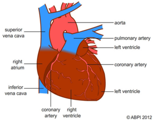
Label Heart Anatomy Diagram Printout - EnchantedLearning.com Oxygen-poor blood enters the right atrium of the heart (via veins called the inferior vena cava and the superior vena cava). The blood is then pumped into the right ventricle and then through the pulmonary artery to the lungs, where the blood is enriched with oxygen (and loses carbon dioxide).
Heart Labelled | Teaching Resources
Human Heart - Anatomy, Functions and Facts about Heart - BYJUS The external structure of the heart has many blood vessels that form a network, with other major vessels emerging from within the structure. The blood vessels typically comprise the following: Veins supply deoxygenated blood to the heart via inferior and superior vena cava, and it eventually drains into the right atrium.
Post a Comment for "38 structure of the heart with labels"